Corneal Endothelial Dysfunction Beyond Fuchs Dystrophy
Understanding Corneal Endothelial Dysfunction
The cornea is the clear dome at the front of your eye, and its innermost layer, called the endothelium, is made up of pump cells that continuously move fluid out of the corneal tissue. When enough of these cells are lost, the cornea swells and vision suffers. Understanding what these cells do, and why they are so difficult to replace, is the foundation of understanding this condition.
Endothelial cells act as a continuous pump, removing excess water from the cornea so it stays thin and transparent. A healthy adult eye has thousands of these cells packed tightly together. The problem is that the human body cannot grow new endothelial cells to replace ones that are lost. Once they are gone, neighboring cells expand to fill the gaps, but over time the pump becomes less efficient.
When endothelial cell loss reaches a critical point, the cornea can no longer stay dry enough to remain clear. Fluid builds up inside the corneal tissue, causing swelling called corneal edema. Early on, this often causes blurry or foggy vision that is worse in the morning and improves as the day goes on. As more cells are lost, the blur becomes permanent, and painful fluid-filled blisters called bullae can form on the corneal surface.
Fuchs dystrophy, a condition in which the endothelial cells gradually deteriorate due to genetics, is the most commonly recognized cause of endothelial dysfunction. However, many other factors can damage these cells, including prior surgeries, infections, trauma, and elevated eye pressure. Each underlying cause may require a different treatment approach, which is why identifying the specific reason for cell loss is an essential part of every evaluation.
Causes of Endothelial Dysfunction Beyond Fuchs
A thorough workup for endothelial dysfunction looks beyond genetics to uncover any contributing factor, because the right treatment depends on understanding the right cause. Our Cornea Specialists are trained to identify and manage the full range of conditions that can damage endothelial cells over a lifetime.
Cataract surgery uses ultrasound energy to break up the cloudy natural lens. This energy can stress nearby endothelial cells, and in cases involving a very dense cataract or a particularly delicate cornea, significant cell loss can follow. When corneal swelling develops months or years after cataract surgery, the condition is called pseudophakic bullous keratopathy. Modern surgical techniques have reduced this risk considerably, but it remains a recognized cause of endothelial damage.
Each eye surgery, including glaucoma procedures, retinal operations, and earlier corneal surgeries, can result in a modest reduction in endothelial cell count. For patients who have undergone several procedures over the years, this cumulative loss can eventually push the cell count below the threshold needed for a clear cornea. Our specialists will check your cell count before planning any future surgery to make sure the cornea can tolerate the procedure safely.
A forceful impact to the eye, whether from a sports injury, a workplace accident, or a motor vehicle collision, can directly damage endothelial cells. In some cases the damage is immediate, but the full effect on cell count may not become apparent for years. If you have experienced significant eye trauma at any point in your life, sharing that history with your eye doctor helps our team understand your baseline and monitor for late-developing changes.
Sustained elevated intraocular pressure, the kind seen with poorly controlled glaucoma, can gradually injure endothelial cells over time. A sudden and severe spike in pressure, as occurs with acute angle-closure glaucoma, can cause rapid and substantial cell loss in a matter of hours. Keeping eye pressure within a safe range through consistent glaucoma management is one of the most effective ways to protect the cells you have.
Certain viral infections can directly target and destroy endothelial cells. Herpes simplex virus affecting the eye is one of the more common infectious causes of endothelial damage. Cytomegalovirus has also been linked to a form of endothelitis, an inflammation of the endothelial layer. Treatment typically involves antiviral medications, which may be given as eye drops, oral pills, or both, tailored to the specific virus involved.
A small number of genetic conditions affect the endothelium independently of Fuchs dystrophy. Posterior polymorphous corneal dystrophy and congenital hereditary endothelial dystrophy are two examples, each with its own pattern of inheritance, appearance, and progression. When a child is born with cloudy corneas, an inherited endothelial dystrophy may be responsible, and specialized evaluation is needed to guide early treatment decisions.
How We Diagnose Endothelial Dysfunction
Accurately diagnosing endothelial dysfunction requires more than a standard eye exam. Our Cornea Specialists use a combination of clinical evaluation and precise diagnostic technology to assess cell health, measure corneal swelling, and identify the underlying cause.
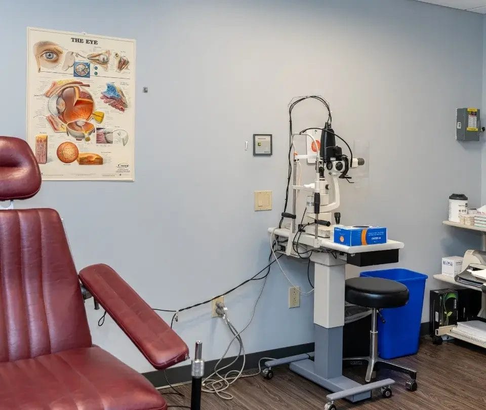
A slit-lamp is a high-powered microscope used to examine the structures of the eye in detail. During this exam, your Cornea Specialist will look for early signs of swelling in the deep layers of the cornea, assess the quality of the endothelial cells using a technique called specular reflection, and evaluate the front segment of the eye for any clues about the cause. Eye pressure, the iris, the lens, and the pupil are all assessed as part of the same visit.
A specular microscope is a specialized camera that photographs and counts the endothelial cells. The test is quick, non-invasive, and provides a precise count of how many cells remain and what their shape and size look like. A healthy adult cornea contains a high density of cells per square millimeter. When the count falls below a critical level, or when the cells become irregular in shape, the risk of corneal swelling rises. Tracking this number over time helps guide treatment timing.
A test called pachymetry measures the thickness of the cornea in microns. A swollen cornea is measurably thicker than a healthy one, making this one of the earliest detectable signs of endothelial trouble. Your Cornea Specialist may include pachymetry at each visit to track whether the cornea is stable, thinning with treatment, or continuing to swell over time.
Some patients benefit from optical coherence tomography, a wide-field scan that creates a detailed cross-sectional image of the corneal layers. This technology can reveal subtle changes in the structure of the endothelium and the tissue immediately above it. If an infectious or systemic cause is suspected, additional testing such as blood work or viral swabs may also be recommended to complete the picture.
Treatment Options for Endothelial Dysfunction
Treatment is matched to the severity of the condition and the underlying cause, with the goal of preserving vision and keeping the corneal surface comfortable. Our approach moves from the least invasive options to surgical repair when necessary, always guided by your symptoms, cell count, and quality of life.
For early-stage swelling, hypertonic saline drops or ointments are prescribed to draw excess fluid out of the cornea. These drops are used multiple times throughout the day and can significantly reduce morning blur in patients with mild dysfunction. Preservative-free lubricating drops and warm compresses can ease surface discomfort. These measures help manage symptoms but do not stop the underlying loss of cells.
When fluid blisters form on the corneal surface, a soft bandage contact lens can provide meaningful pain relief. The lens creates a smooth barrier that protects the sensitive corneal nerves from the friction of blinking. It is typically worn alongside antibiotic drops to prevent infection. A bandage lens is usually a temporary measure, offering comfort while a longer-term plan, including surgery, is being arranged.
For moderate to advanced cases, the most effective and lasting treatment is an endothelial transplant. This procedure replaces only the damaged inner layer with healthy donor tissue, leaving the rest of the cornea intact. There are two primary approaches used at our practice.
- DMEK (Descemet Membrane Endothelial Keratoplasty) transplants the thinnest possible layer of donor tissue and typically offers the sharpest final vision.
- DSAEK (Descemet Stripping Automated Endothelial Keratoplasty) uses a slightly thicker donor layer and may be preferred in eyes with complex anatomy or prior surgeries.
Our Cornea Specialists, including Dr. Jane Cook, Dr. Elliot Perlman, and Dr. Christopher Newton, perform both procedures and will recommend the approach best suited to your eye. Most patients experience meaningful vision improvement within weeks of surgery.
In cases where the entire cornea has been severely scarred or damaged, a full-thickness transplant called penetrating keratoplasty may be necessary. This replaces the complete cornea with donor tissue and involves a longer recovery than endothelial keratoplasty. For patients whose eye is blind and painful, and where vision restoration is not the primary goal, comfort-focused options such as a conjunctival tissue cover may be used to relieve pain without a full transplant.

Frequently Asked Questions
Here are answers to some of the questions our patients ask most often about endothelial dysfunction and the care options available to them.
General eye doctors play an important role in early detection and ongoing monitoring, but when endothelial cell counts are declining significantly or vision is noticeably affected, a consultation with a Cornea Specialist is strongly advisable. Complex cases involving prior surgeries, infections, or rare inherited conditions benefit from the level of subspecialty expertise and surgical capability that a dedicated cornea program provides. Our Cornea Specialists work closely with referring optometrists and general ophthalmologists to coordinate seamless care.
Any new or worsening blur, especially blur that is present in the morning and improves over a few hours, is worth reporting to your eye doctor. If the blur is lasting all day, if your eye is red and painful, or if you notice blisters or a gritty sensation on the surface of the eye, you should seek evaluation promptly. Sudden changes in vision after any previous eye surgery are also a reason to call sooner rather than later.
Not necessarily. In many situations, cataract surgery and an endothelial transplant can be performed together in a single combined procedure, which reduces the number of surgeries and may lead to better visual outcomes. When they need to be staged separately, our team plans the sequence based on what is safest for your cornea and most likely to give you the clearest vision. The right approach depends on your individual anatomy, cell count, and the severity of your cataract.
Donor tissue can, in some cases, be rejected by the immune system, a process called graft rejection. The risk is manageable and monitoring is ongoing. Signs of rejection include sudden redness, light sensitivity, decreased vision, or pain, and any of these symptoms after a transplant should prompt an urgent call to our office. When caught early, rejection episodes can often be treated successfully with medicated drops. Your Cornea Specialist will explain the warning signs clearly before you leave the surgical suite.
Yes, in some cases. The most important protective steps are controlling eye pressure if you have glaucoma, treating eye infections promptly and completely, wearing protective eyewear in high-risk settings, and keeping all scheduled eye appointments so that cell loss is tracked consistently. Hypertonic drops may also slow the functional impact of cell loss in some patients, though they do not prevent the underlying decline. Early detection gives your care team the most time to plan ahead with you.
Cell cultivation and regenerative approaches for the corneal endothelium are active areas of research, and some early clinical trials are underway in specialized centers. However, these options are not yet available as standard care for most patients. The current gold standard for significant endothelial cell failure remains transplantation with healthy donor tissue. Our team stays current with emerging treatments and can discuss whether any evolving options may be relevant to your situation in the future.
See Our Cornea Specialists at Rhode Island Eye Institute
Whether your endothelial dysfunction stems from a prior surgery, a viral infection, an inherited condition, or a cause still being identified, our fellowship-trained Cornea Specialists are here to help you understand what is happening and what to do about it. Rhode Island Eye Institute brings together surgical expertise, advanced diagnostic technology, and integrated optometric co-management under one roof, so your care is coordinated from your first visit through every follow-up. We welcome patients from across the region and are ready to partner with you in protecting your cornea and your vision.