Corneal Graft Failure: What It Means and What Comes Next
Understanding Corneal Graft Failure
Corneal graft failure is the lasting loss of clarity or function in a transplanted cornea. It is an important distinction from rejection, and knowing the difference helps guide the right response and the right treatment.
Rejection is an immune response in which the body attacks the donated tissue. When caught early and treated with steroid eye drops, many rejection episodes can be reversed before permanent damage occurs. Graft failure, by contrast, is the point at which the cornea no longer clears or functions, even with treatment. Every untreated or repeat rejection episode brings the graft closer to failure.
A failed corneal graft cannot restore clear sight on its own. The cornea may remain cloudy or swollen, and glasses or contact lenses cannot fully correct the blurred vision that results. Many patients are candidates for a new transplant or an alternative surgical procedure, and your Cornea Specialist will help map out the most appropriate path forward.
Failure in the early months after transplant is uncommon when surgery goes well. Over the long term, the risk increases as the specialized inner pump cells of the cornea, called endothelial cells, gradually decline in number. Most late-stage failures result from this slow, cumulative pump cell loss. Careful follow-up helps detect early changes before vision is significantly affected.
Ongoing care with a Cornea Specialist is not optional after a transplant. Anti-rejection eye drops are typically continued for years, sometimes at a low maintenance dose indefinitely. Routine visits allow your care team to measure pump cell counts, monitor eye pressure, and assess the graft surface before small problems become serious ones.
What Causes Corneal Graft Failure
Graft failure can develop through several different pathways, some apparent within days of surgery and others unfolding over many years. Identifying the cause is the first step in choosing the right next treatment.
Primary graft failure presents as persistent corneal swelling and cloudiness beginning in the first week after surgery. It typically reflects inadequate pump cell function in the donor tissue from the outset, rather than anything related to the patient's immune response. A repeat transplant is usually required. Your surgeon will evaluate the situation within the first weeks and discuss the available options.
A single rejection episode caught and treated early often resolves without lasting damage. Repeated episodes, or episodes that go unrecognized and untreated, damage the pump cells with each occurrence. Over time, the cumulative loss of these cells reaches a point where the graft can no longer remove fluid from the cornea, and it becomes permanently cloudy.
Late pump cell loss is the most common cause of long-term failure following both full-thickness and partial-thickness transplants. Pump cells decline gradually in every graft, even a healthy and stable one. Factors that accelerate this loss include prior eye surgeries, elevated eye pressure, glaucoma, and previous rejection episodes. When the reserve of surviving cells falls too low, the cornea loses its ability to stay clear.
Infection of a transplanted cornea is a serious threat to graft clarity. Herpes simplex virus can reactivate in graft tissue. Bacterial or fungal infections, sometimes associated with a loose suture or a break in the surface, can also develop. These situations require same-day evaluation and culture-directed treatment to prevent irreversible damage.
A corneal surface wound that does not heal, called a persistent epithelial defect, can threaten the health of the graft beneath it. Elevated eye pressure (glaucoma) directly damages endothelial pump cells and raises the risk of failure over time. Managing both the surface and pressure with drops, lubricants, or other treatments is an important part of protecting any transplanted cornea.
Warning Signs That Need Same-Day Care
Many rejection episodes and early graft-threatening problems can be reversed when treated promptly. Recognizing the warning signs and acting the same day you notice them gives the graft its best chance. Do not wait for a scheduled appointment if any of these symptoms appear.
A shift from clear to hazy vision in the eye that received the transplant is one of the most important warning signs to know. It may develop over hours or over several days. It should never be attributed to tiredness or dry eyes without a clinical evaluation. Contact the cornea care team for a same-day visit.
A graft that becomes red, particularly with a ring of redness around its edge, may be experiencing an immune attack. Pain ranging from mild to sharp can accompany this finding. Both redness and pain following transplant surgery warrant prompt evaluation, not watchful waiting.
New sensitivity to light, especially when combined with redness or tearing, is a recognized warning sign of graft stress or rejection. Bright indoor or outdoor light may feel unusually harsh. These symptoms often indicate that a closer examination is needed right away.
A noticeable drop in how sharply you can see, or a sudden change after a long period of stable vision, is urgent. A decline of two lines or more on the eye chart is a clinical red flag. Early intervention at this stage frequently makes the difference between a reversible event and permanent graft failure.
How We Evaluate a Failing Graft
When a patient comes in with signs of graft distress, our Cornea Specialists use a combination of clinical examination and specialized testing to understand exactly what is happening. The goal is to determine whether the process can be reversed and what intervention is most appropriate.
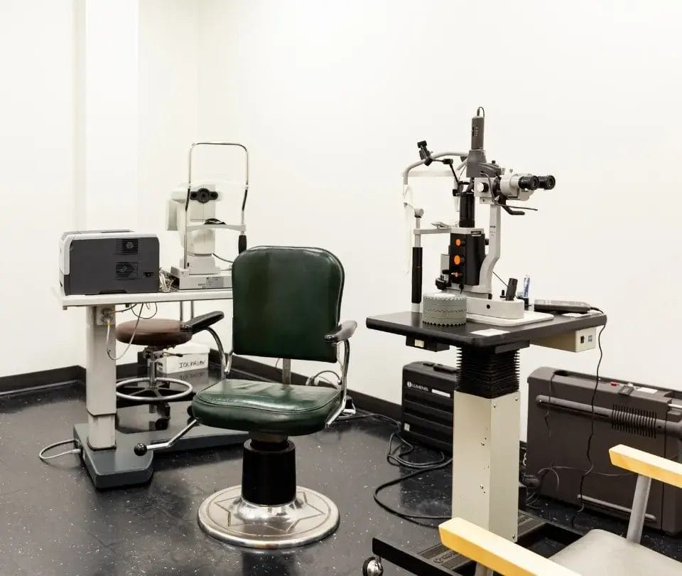
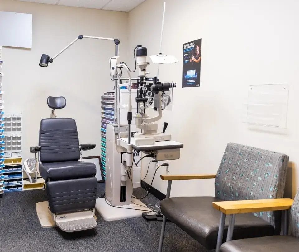
A slit lamp is a microscope paired with a focused beam of light that allows each layer of the cornea to be examined in detail. Your Cornea Specialist uses it to identify swelling, pump cell changes, surface defects, and signs of infection or immune activity. This examination forms the foundation of every graft evaluation.
Pachymetry is a quick, painless test that measures corneal thickness. A graft that is swelling with fluid becomes measurably thicker than its normal baseline. Specular microscopy counts and maps the remaining pump cells in the graft. Low pump cell counts confirm late failure from endothelial cell loss and help guide decisions about timing and type of repeat surgery.
Eye pressure is checked at every visit because elevated pressure is both a cause and a consequence of graft problems. A newly elevated pressure may help explain why a graft is failing. Drops or laser treatment can reduce pressure and protect any remaining healthy pump cells while next steps are planned.
When infection is suspected, a corneal scraping is collected and sent for laboratory analysis. Testing may include a Gram stain, culture for bacteria and fungus, and a PCR test to detect herpes simplex virus DNA. These results direct the most targeted and effective antimicrobial treatment available.

Treatment Options After Graft Failure
The right treatment depends on the cause of failure, the overall health of the eye, and how much vision has been affected. Our cornea team reviews each case individually to match the procedure to the patient.
A repeat penetrating keratoplasty, sometimes called repeat PK, replaces the entire thickness of the cornea with a new donor graft. Repeat grafts carry a higher risk of rejection and failure compared to a first transplant, which makes careful patient selection and consistent use of anti-rejection drops especially important. Recovery takes several months, and vision improvement is gradual.
When only the inner pump cell layer of the cornea has failed, an endothelial keratoplasty can be placed beneath an existing graft or alongside other transplant strategies. DMEK (Descemet membrane endothelial keratoplasty) and DSAEK (Descemet stripping automated endothelial keratoplasty) are the two primary techniques. Both replace only the damaged inner layer, leaving healthy tissue undisturbed, and recovery is typically faster than full-thickness surgery. Dr. Jane Cook, Dr. Elliot Perlman, and Dr. Christopher Newton each perform endothelial keratoplasty procedures.
Deep anterior lamellar keratoplasty, or DALK, replaces only the front layers of the cornea while preserving the patient's own healthy inner pump cell layer. Because the patient's endothelial cells remain in place, the risk of rejection is substantially lower than with a full-thickness transplant. DALK is best suited to cases where the outer or middle layers have failed but the inner layer remains functional.
A keratoprosthesis is an artificial corneal device used when conventional transplantation has failed multiple times and the eye is no longer a suitable candidate for donor tissue. It can restore meaningful functional vision in otherwise difficult-to-treat situations. Lifelong monitoring by a Cornea Specialist is essential for patients with a keratoprosthesis in place.
While surgical planning is underway, lubricating eye drops and bandage contact lenses can improve comfort and help protect the graft surface. Pressure-lowering drops preserve any remaining pump cell function. Our team will establish a clear bridge care plan so that no patient is left without guidance in the interval before their next procedure.
Frequently Asked Questions
These questions reflect common concerns that arise after a graft becomes cloudy or a rejection episode occurs. Each answer is intended to help you make more informed decisions in conversation with your Cornea Specialist.
Prompt treatment with steroid eye drops does reverse many rejection episodes before they cause permanent damage to the pump cells. The key word is prompt. Waiting even a few days can allow enough cell loss to occur that recovery is incomplete. If you have any of the warning signs described above, contacting the cornea office the same day, rather than waiting for a scheduled appointment, gives the graft its best chance of survival.
Yes. Eyes with prior surgeries, elevated eye pressure, a history of herpes simplex disease, or previous rejection episodes carry a higher long-term failure risk. Patients who received a full-thickness transplant, rather than a partial-thickness one, also tend to lose pump cells more quickly over time. Knowing your individual risk profile helps your Cornea Specialist tailor your follow-up schedule and your drop regimen to reduce that risk.
Most patients continue topical steroid drops for years after a transplant, and some remain on a low-dose maintenance regimen indefinitely. Stopping drops prematurely is one of the leading preventable causes of late rejection. If cost, availability, or side effects are concerns, discuss them directly with your surgeon so that an alternative plan can be arranged. Never discontinue drops on your own without guidance.
Pump cells decline gradually throughout the life of any graft. Even a long-stable, well-functioning transplant reaches a tipping point if the cell count falls low enough. This is called late endothelial failure, and it can appear after a decade or more of clear vision. It does not necessarily mean anything went wrong; it reflects the natural biology of transplanted tissue. A timely evaluation can determine whether a repeat procedure is appropriate and which type is the best fit for your eye.
Traveling is generally safe once your surgeon has cleared you after surgery. For ongoing post-transplant care, carry your eye drops in your carry-on bag and bring enough supply to cover the full trip, plus extra. Cabin air is typically dry, so lubricating drops are helpful during flights. Make sure you have your surgeon's contact information available in case symptoms appear while you are away from home.
Most major insurers in the United States cover corneal transplantation, including repeat procedures, when medical necessity is established. Pre-authorization is often required before a repeat surgery is scheduled. Our team can assist in verifying your specific coverage and preparing the documentation needed before your procedure is booked.
Schedule a Graft Review at Rhode Island Eye Institute
If your transplanted cornea has become cloudy, if warning signs have appeared, or if it has simply been a while since your last pump cell count was checked, we are here to help. At Rhode Island Eye Institute, our fellowship-trained Cornea Specialists, including Dr. Jane Cook, Dr. Elliot Perlman, and Dr. Christopher Newton, bring deep experience in evaluating and managing failed and at-risk grafts. We welcome patients from across Rhode Island and Southern Massachusetts and are committed to giving every transplant patient the careful, ongoing attention their vision deserves. Contact us today to schedule your corneal graft evaluation.