Eye Floaters and Flashes
When Floaters and Flashes Become an Emergency
Most floaters are not dangerous, but certain symptoms are warning signs that need same-day attention. Knowing the difference between a routine floater and an emergency can protect your vision and, in some cases, prevent permanent vision loss.
A sudden burst of new floaters, a rapid increase in flashes of light, or a dark curtain or shadow moving across your vision can all signal a retinal tear or retinal detachment. The retina is the light-sensitive tissue lining the back of your eye, and when it tears or detaches, your vision is at serious risk.
The key word is sudden. Floaters you have had for years are typically not a concern. A new shower of spots, new flashes, or a shadow in your side vision that appeared out of nowhere should be treated as an emergency.
Contact your eye doctor immediately if you experience any of these warning signs. If you cannot reach your eye doctor the same day, go to an emergency room without delay. Do not wait to see if the symptoms resolve on their own, as retinal detachment can lead to permanent vision loss without prompt treatment.
While you are waiting to be seen, avoid heavy lifting, bending at the waist, or any jarring physical activity that could worsen a retinal tear. Take note of which eye is affected and when you first noticed the symptoms, since your doctor will need these details during your evaluation.
A retinal tear that is caught early can often be sealed with an in-office laser procedure or a freezing treatment called cryopexy, which prevents a full detachment from developing. Once the retina detaches, surgery is required and the outcome depends heavily on how quickly treatment is received.
If the central part of the retina, called the macula, becomes involved in the detachment, the risk of lasting central vision loss increases significantly. Patients who receive treatment within the first day or two generally have a better chance at visual recovery than those who wait.

What Causes Floaters and Flashes
Floaters and flashes have several causes, most of which are related to natural changes inside the eye as we age. Understanding what is happening inside your eye can help you recognize what is normal and what warrants a call to your doctor.
The most common cause of new floaters in adults, especially those over 50, is a condition called posterior vitreous detachment, or PVD. The vitreous is a clear, gel-like substance that fills the inside of your eye. As you get older, this gel naturally shrinks and pulls away from the retina, a process that is a normal part of aging.
As the vitreous separates, clumps and strands of gel fiber cast shadows on the retina, which you perceive as floaters. The tugging motion of the vitreous against the retina also produces the flashes of light many patients describe. PVD itself is not dangerous, but the pulling force can sometimes create a retinal tear, which is why new floaters always need a thorough retinal examination.
Even without a full PVD, the vitreous gel naturally breaks down over time, forming small strands and clumps that drift through your field of vision. You are most likely to notice these floaters when looking at a bright sky, a white wall, or a computer screen.
These age-related floaters tend to become less noticeable over weeks to months. They settle lower in the eye and your brain gradually learns to filter them out, which is why older floaters often bother you much less than newer ones.
While floaters can appear at any age, certain factors increase the likelihood of developing PVD and related symptoms earlier. These include being over age 50, having high myopia (nearsightedness), a history of eye surgery, or a prior eye injury.
- People with high myopia tend to develop PVD sooner because their eyes are physically longer, which places more tension on the vitreous gel
- Cataract surgery can change the structure inside the eye in a way that accelerates vitreous changes
- If you have had a PVD in one eye, there is a meaningful chance of developing one in the other eye within a few years
Letting your eye doctor know about these risk factors helps them monitor your retinal health more closely over time.
How We Diagnose and Treat Floaters
When you come in with new floaters or flashes, our first priority is to make sure your retina is healthy and intact. From there, we help you understand your options based on what we find.
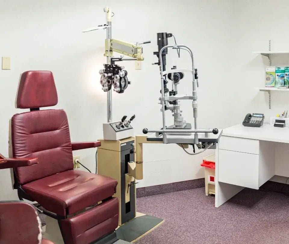
Your provider will dilate your pupils and examine your entire retina using a bright light and magnifying lenses. This dilated eye exam allows us to check for retinal tears, areas of thinning, holes, and any signs of detachment. In some cases, if the vitreous is too cloudy to see through clearly, we may use ocular ultrasound to get a complete picture.
One exam may not be enough to give you full assurance. Because retinal tears can develop days to weeks after a PVD begins, your doctor may ask you to return for a follow-up visit in four to six weeks, even if the first exam looks normal. It is important to keep that appointment even if your symptoms have improved.
When your exam confirms that your floaters are benign, meaning there are no tears or other retinal problems, no treatment is needed. Over time, most people find that floaters become far less bothersome as they settle in the eye and the brain adapts to their presence.
If a floater drifts into your line of sight during a task, gently moving your eyes up and down or side to side can shift it out of your direct vision. Most people adjust to benign floaters within a few months without any medical intervention.
In a small number of patients, floaters are large or numerous enough to significantly interfere with reading, driving, or screen-based work. In these situations, your doctor may discuss two procedural options.
- Vitrectomy is a surgical procedure that removes the vitreous gel and its floaters, replacing it with a clear saline solution. It is effective but carries risks including cataracts, retinal tears, and infection, so it is reserved for cases with major functional impact
- Laser vitreolysis uses concentrated laser pulses to break large floaters into smaller, less visible fragments. It is less invasive than vitrectomy but is not suitable for all floater types or locations
Your doctor will evaluate the size, location, and nature of your floaters to determine whether either option is appropriate for your specific situation.
Frequently Asked Questions
These answers address common questions patients ask when they first notice floaters or flashes, with a focus on helping you decide when to act and what to expect.
Floaters rarely disappear entirely, but they do become much less noticeable for most patients over time. As they settle lower in the eye and your visual system adapts, the brain begins to filter them out of your conscious awareness. New floaters from a recent PVD tend to be the most bothersome at first, but the majority of patients report significant improvement within a few months without any treatment.
Brief flashes when you move your eyes in darkness can be a normal part of an active PVD and are not automatically a cause for alarm. However, frequent or persistent flashes, particularly when accompanied by new floaters, should be evaluated promptly. The concern is whether the flashes indicate harmless vitreous movement or a retinal tear that requires treatment, and only a dilated retinal exam can make that distinction.
If you experience a sudden shower of new floaters, new flashes, or any shadow or curtain in your vision, and you cannot reach your eye doctor the same day, going to the emergency room is the right decision. ER physicians can perform an initial evaluation and connect you with an ophthalmologist for a detailed retinal exam. Speed matters when a retinal tear or detachment is possible, and it is always better to be seen and reassured than to wait.
Routine exercise does not cause floaters in a healthy eye. However, if you have recently been diagnosed with a PVD or a retinal tear, your doctor may advise you to temporarily avoid heavy lifting, contact sports, or activities involving significant jarring or straining. These movements can potentially increase tension on a partially separated vitreous. Ask your doctor about specific activity guidelines if you have a recent PVD diagnosis before returning to your normal routine.
For the majority of patients, floaters are a benign sign of normal aging inside the eye and do not indicate a serious underlying condition. That said, any new floaters still warrant a dilated retinal exam to rule out retinal tears and other issues that could threaten your vision. Once your retina has been examined and confirmed to be healthy, you can feel confident that your floaters are not a danger to your sight.
After your initial exam, your doctor will set a follow-up schedule based on your specific situation, typically within four to six weeks of a new PVD. Beyond that, routine comprehensive eye exams are important for anyone with a history of vitreous changes or retinal risk factors. If you notice any new sudden changes in your floaters or flashes between scheduled visits, contact us right away rather than waiting for your next appointment.

See Our Team for Expert Retinal Care
At Rhode Island Eye Institute, our retina specialists have the advanced training and technology to evaluate your symptoms thoroughly and provide guidance you can trust. Whether you need an urgent evaluation or a routine floater check, we are here to protect your vision and give you peace of mind. We welcome patients from across Rhode Island and are committed to providing the responsive, expert care your eyes deserve.