
Glaucoma After Cataract Surgery
Why Eye Pressure Can Rise After Cataract Surgery
During cataract surgery, we remove the cloudy natural lens and replace it with a clear artificial one. This process can temporarily change how fluid flows through your eye, and in some patients, that change causes pressure to build up. Most pressure rises after surgery are short-lived, but some can develop into a longer-term condition that needs ongoing care.
The most common pressure-related issue after cataract surgery is a temporary spike, not true glaucoma. These spikes typically occur in the first day or two and usually resolve quickly with treatment.
- Retained viscoelastic gel, a clear fluid used during surgery to protect the eye's structures, left behind after the procedure
- Steroid eye drops prescribed after surgery causing pressure to rise in sensitive patients
- Inflammation or swelling in the eye's drainage system
- A small amount of bleeding or red blood cells inside the eye
- Microscopic lens fragments triggering inflammation
A single elevated pressure reading does not mean you have glaucoma. Our eye doctor will assess the full picture before drawing any conclusions.
Less commonly, patients develop sustained high pressure that causes lasting changes to the optic nerve or visual field. This is called secondary glaucoma, meaning it has a specific underlying cause related to surgery or healing.
Open-angle glaucoma can develop when the drainage system becomes less efficient even though the drainage angle appears open. Angle-closure glaucoma can occur when the drainage pathway becomes physically blocked. A rare condition called malignant glaucoma, or aqueous misdirection, happens when fluid is redirected backward in the eye, causing the front chamber to shallow and pressure to spike. This requires urgent treatment.
Several additional processes can raise eye pressure after cataract surgery. UGH syndrome (uveitis, glaucoma, and hyphema) occurs when a poorly positioned lens implant irritates the iris and causes ongoing inflammation and bleeding. Scarring called peripheral anterior synechiae can close off the drainage angle over time, and ghost cell glaucoma can result from old blood cells migrating into the drainage system after a vitreous hemorrhage. Our eye doctor will consider all of these possibilities when evaluating your situation.
The eye continuously produces and drains a fluid called aqueous humor to maintain healthy pressure. Surgery can temporarily disrupt this balance in several ways.
- Surgical instruments and irrigating fluid can cause transient swelling in drainage channels
- Lens fragments or residual gel may partially block normal outflow
- The position of the new artificial lens can alter the geometry of the drainage angle
- Healing tissue can form in areas that affect how fluid exits the eye

Who Is at Higher Risk
Anyone can experience a temporary pressure rise after cataract surgery, but certain factors make it more likely. Knowing your risk level helps us plan appropriate monitoring and catch problems early.
If you already have glaucoma or borderline high eye pressure before surgery, your risk of pressure problems afterward is greater. Other conditions that increase risk include a history of eye inflammation, narrow drainage angles, or a prior eye injury.
- Diagnosed glaucoma or ocular hypertension before surgery
- Narrow angles or a shallow anterior chamber, the front compartment of the eye
- Prior episodes of uveitis, or inflammation inside the eye
- Pseudoexfoliation syndrome, a condition where flaky material builds up in the eye's drainage structures
- Previous eye surgery or trauma
The vast majority of cataract surgeries proceed smoothly, but certain intraoperative events can raise the likelihood of elevated pressure afterward. A torn capsule, bleeding inside the eye, or difficulty placing the new lens can all create conditions that affect drainage. If any of these occur during your procedure, our eye doctor will monitor you more closely than usual during recovery.
Steroid eye drops are a standard and important part of recovery after cataract surgery. However, some patients experience steroid-induced glaucoma, in which the anti-inflammatory drops themselves cause pressure to rise. This is more likely with prolonged or high-dose steroid use. Inflammation can also raise pressure independently by flooding the drainage system with inflammatory cells and proteins that clog normal outflow.
As we age, the drainage system inside the eye naturally becomes less efficient. This means older adults may have less reserve to handle the additional stress that surgery places on the eye's fluid balance. Systemic conditions that affect healing, such as diabetes, can also contribute to prolonged inflammation and elevated pressure.
- Advanced age with reduced natural drainage function
- Diabetes or conditions that slow healing and increase inflammation
- A family history of glaucoma
- High myopia, or significant nearsightedness, associated with longer eye length
Warning Signs and When to Seek Care
Knowing which symptoms to watch for after cataract surgery can make a meaningful difference in protecting your vision. Some signs are mild and simply warrant a timely call to our office, while others need immediate attention.
In the days and weeks following surgery, mild discomfort, a feeling of fullness or pressure in the eye, slight blurriness, or halos around lights can be early clues that pressure is rising. These symptoms do not always indicate glaucoma, but they should be reported to our office so we can evaluate you promptly. Early detection gives us the best chance to prevent lasting damage.
Certain symptoms signal a serious or rapidly worsening problem that needs urgent care. Do not wait to see if these resolve on their own, and do not press on your eye to check it yourself, as this can cause harm after surgery.
- Sudden sharp or throbbing pain in or around the eye
- Severe headache accompanied by eye discomfort
- Nausea or vomiting alongside eye symptoms
- Rapid or sudden loss of vision
- A curtain, shadow, or dark area blocking part of your vision
- Extreme redness with intense pain
Some fluctuation in vision is normal during early recovery, but certain changes should prompt a same-day call to our office. A sudden drop in vision, new blind spots, persistent cloudiness that is getting worse rather than better, dimmer colors, or narrowing side vision can all indicate elevated pressure or another complication. A shadow or curtain in your vision can also signal retinal detachment, which is a separate urgent condition that always requires emergency evaluation.
For mild discomfort, slight blurriness, or minor vision changes during normal business hours, call our office so we can arrange a prompt examination. We can typically see you the same day or within 24 hours to check your pressure and assess what is happening.
For severe pain, sudden vision loss, or intense headache with nausea, contact your surgeon or our on-call ophthalmology service right away. If symptoms occur after hours and you cannot reach our team promptly, go to the nearest emergency department and ask for urgent eye evaluation. Keep in mind that not all emergency rooms are fully equipped to manage eye pressure emergencies, so reaching your eye care team directly is always the first priority.
How We Diagnose Post-Surgical Glaucoma
Accurate diagnosis is the foundation of effective treatment. We use a combination of measurements, specialized examinations, and imaging to understand exactly what is happening inside your eye.
The first step is measuring your intraocular pressure, often abbreviated as IOP, using an instrument called a tonometer. The test is quick and painless and gives us a number to compare with your pre-surgery baseline when available. Because eye pressure naturally fluctuates throughout the day, we may measure it at different times or on different days to determine whether you have a temporary spike or a persistent problem.
To understand why pressure may be elevated, we examine the drainage angle where fluid exits the eye. We use a technique called gonioscopy, in which a small mirrored contact lens is placed on your numbed eye, allowing us to see whether the angle is open, narrow, or blocked by inflammation, scar tissue, or debris. This examination directly guides our treatment choices.
- Numbing drops are applied before the lens is placed, so the procedure is comfortable
- The mirrored lens shows us the drainage structures that are otherwise not visible
- We look for blockages, scarring, angle closure, and any abnormal tissue
- Results help us match your treatment to the specific cause of your pressure elevation
Because sustained high pressure can damage the optic nerve, we examine it carefully at every visit. We use magnifying lenses and a microscope to look at the nerve head for signs of damage such as increased cupping, which refers to enlargement of the central depression in the nerve, or thinning of nerve tissue. We also use optical coherence tomography, or OCT, to create detailed cross-sectional images of the optic nerve and surrounding layers. Color stereoscopic optic nerve photography provides a visual record we can compare over time to detect subtle changes early.
Glaucoma often causes gradual loss of peripheral, or side, vision before central vision is affected. Visual field testing maps out your full range of vision by measuring how reliably you detect light flashes at different positions while you focus on a central target. Comparing results across multiple visits tells us whether glaucoma is affecting your sight and whether treatment is preventing further progression.

Treatment Options for Post-Surgical Glaucoma
Treatment depends on the specific cause and severity of your pressure elevation. Our glaucoma specialist, Dr. Sarah Anis, tailors care to each patient's unique anatomy, surgical history, and degree of optic nerve involvement.
When a specific cause is identified, addressing it directly is the first priority. Retained viscoelastic gel may be cleared with a washout procedure or managed with medication. Significant lens fragments causing inflammation may require surgical removal. Pupillary block or angle closure may be treated with a laser peripheral iridotomy, a procedure that creates a small opening in the iris to restore normal fluid flow. When malignant glaucoma is diagnosed, treatment includes cycloplegic drops to relax the eye, medications to reduce fluid production, and sometimes laser or surgical intervention to correct fluid misdirection.
Most patients with elevated pressure after cataract surgery respond well to medicated eye drops that either reduce the amount of fluid the eye produces or improve how well it drains. Common classes include prostaglandin analogs, beta blockers, alpha agonists, and carbonic anhydrase inhibitors. Not every drop is appropriate for every patient. For example, beta blockers are avoided in patients with certain breathing or heart conditions, and prostaglandin analogs are timed carefully based on your inflammation status and risk of macular swelling. Our eye doctor will select the combination that is safest and most effective for you.
When pressure rises sharply in the hours or days after surgery, oral medications such as acetazolamide can quickly reduce fluid production and bring pressure down while we identify and address the underlying cause. These medications are typically used short-term and can cause side effects including tingling in the hands and feet, altered taste, or increased urination. We monitor you closely and adjust therapy as your pressure stabilizes.
If drops alone are not sufficient, laser trabeculoplasty uses a focused beam of light to open the trabecular meshwork and help fluid leave the eye more efficiently. This is an option for open-angle mechanisms and is generally considered once your eye has healed and inflammation has settled. It is not used for angle-closure problems, pupillary block, or acute inflammatory pressure spikes.
- Numbing drops are used so the procedure is comfortable
- The laser targets specific areas of the drainage tissue in a brief in-office visit
- Pressure may improve gradually over several weeks after treatment
- Many patients still use drops afterward, though sometimes fewer are needed
When drops and laser treatment do not bring pressure down to a safe level, minimally invasive glaucoma surgery, or MIGS, offers a surgical option with a relatively favorable recovery profile. MIGS procedures use micro-stent implants to create or enhance drainage pathways inside the eye. Dr. Sarah Anis performs these procedures and can combine them with cataract surgery when both conditions are present, allowing patients to address both issues in a single operation.
In cases where MIGS is not sufficient, more involved surgeries such as trabeculectomy or tube shunt implantation may be recommended. Trabeculectomy creates a new drainage pathway through the eye wall, while a tube shunt implants a small device that channels fluid out of the eye and reduces pressure. The right choice depends on your specific type of glaucoma, the anatomy of your eye, and how well earlier treatments have worked. Our eye doctor will review each option carefully with you so you can make an informed decision.
When steroids are contributing to elevated pressure, we may taper the dose, switch to a lower-potency anti-inflammatory medication, or add pressure-lowering drops while continuing necessary steroid therapy. When inflammation itself is clogging the drainage system, adequate anti-inflammatory treatment is essential alongside pressure-lowering medications. Do not reduce or stop your steroid drops on your own without guidance from our office, as doing so can worsen inflammation and cause other complications. We will always find the safest balance between controlling inflammation and protecting your drainage system.
Recovery and Long-Term Monitoring
Healing from cataract surgery and any additional glaucoma treatment takes time, and ongoing monitoring is essential to catch changes before they affect your sight. We stay closely involved throughout your recovery and beyond.
Your vision will likely continue to improve over several weeks as swelling decreases and your eye adjusts to its new lens. Some mild discomfort, light sensitivity, or day-to-day fluctuation in vision is normal during this period. If these symptoms worsen or new ones develop, contact our office right away rather than waiting for your next scheduled visit.
We typically schedule follow-up visits at one day, one week, one month, and three months after surgery, though your schedule may vary depending on your individual situation. If your pressure is elevated or we suspect glaucoma, you may need more frequent check-ins, sometimes same-day or next-day rechecks, until your pressure is consistently stable.
- Early visits focus on healing, pressure monitoring, and drop adjustments
- Later visits evaluate your optic nerve health and visual field
- Long-term monitoring continues even after full healing is confirmed
Following your post-operative instructions carefully gives your eye the best chance to heal well and keeps pressure stable. Use all prescribed drops on schedule, even when your eye feels comfortable, because consistent use is essential for pressure control. Avoid rubbing or pressing on your eye, wear any protective shield or glasses as directed, avoid heavy lifting or strenuous activity for the first few weeks, and keep every follow-up appointment. If you have trouble using your drops or run out before your next visit, let us know right away.
Even after full recovery from surgery, some patients require ongoing treatment to maintain healthy eye pressure. Others find that pressure stabilizes and drops can eventually be reduced or discontinued. Regular eye exams remain essential regardless of how well things are going, as glaucoma can change slowly and silently. Continued monitoring of your pressure, optic nerve, and visual field gives us the best opportunity to catch any shift early and adjust your care accordingly.
Frequently Asked Questions
These answers address common questions patients have after learning their eye pressure is elevated following cataract surgery.
Not necessarily, particularly when it is detected and treated early. Most patients who receive timely care maintain excellent vision. However, if pressure remains very high for an extended period before treatment begins, some optic nerve damage can be permanent. This is why attending every post-operative visit and reporting symptoms promptly matters so much, even when you feel fine overall.
Yes. While many cases arise in the first days to weeks, delayed pressure elevation is possible months or even years later. Gradual scarring of the drainage angle, slow lens position changes, or factors entirely unrelated to the original surgery can all cause late-onset pressure rise. Routine annual eye exams are the most reliable way to detect these delayed changes before vision is affected.
They share the same core risk, which is damage to the optic nerve from elevated pressure, but the causes are different. Post-surgical glaucoma has specific triggers tied to the healing process, inflammation, or structural changes from surgery. Treatment principles overlap, but the sequence and selection of therapies are tailored to what caused the pressure rise in your particular case. Your care plan may differ significantly from someone with primary open-angle glaucoma who never had surgery.
No. Cataract surgery is highly effective at restoring clear vision, and the lens replacement itself typically functions well regardless of what happens with eye pressure. Developing a secondary pressure issue is a separate matter from how well your new lens is working. Most patients with post-surgical glaucoma continue to enjoy the visual improvement from their cataract procedure while managing their pressure through medication, laser treatment, or surgery.
It depends on the cause and your eye's response to therapy. Some patients need treatment for only a short period while the underlying cause resolves. Others, particularly those with pre-existing glaucoma or significant optic nerve involvement, require long-term management. We reassess your treatment plan regularly and aim to use the least intervention necessary to keep your pressure at a safe level while protecting your vision over time.
If you have risk factors such as narrow angles, pseudoexfoliation syndrome, or a history of high pressure, we take those into account before and during surgery on your second eye. Knowing your history allows us to plan more carefully, monitor more closely, and intervene earlier if pressure rises. Having pressure problems in one eye does not guarantee the same will happen in the other, but it does raise our level of vigilance.
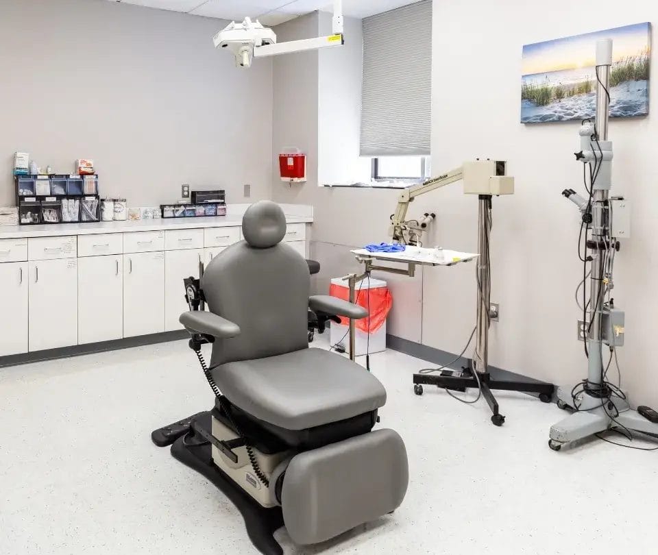
Glaucoma Care at Rhode Island Eye Institute
If you have had cataract surgery and have concerns about your eye pressure, vision, or recovery, our team is here to help. At Rhode Island Eye Institute, our glaucoma specialists have the training, technology, and experience to accurately diagnose and treat the full range of pressure-related conditions that can arise after surgery. We are committed to protecting your vision for the long term and will work with you every step of the way.